Home » Without Label » Deigram Of Outside Leg Muscles - Cardiovascular System Of The Leg And Foot / Reflexes help to maintain proper muscle tone, balance, and responsiveness of the legs and feet to stimuli such as stepping on a sharp object.
Deigram Of Outside Leg Muscles - Cardiovascular System Of The Leg And Foot / Reflexes help to maintain proper muscle tone, balance, and responsiveness of the legs and feet to stimuli such as stepping on a sharp object.
Deigram Of Outside Leg Muscles - Cardiovascular System Of The Leg And Foot / Reflexes help to maintain proper muscle tone, balance, and responsiveness of the legs and feet to stimuli such as stepping on a sharp object.. Groin muscles help support the hip joint. It contains the peroneus longus and peroneus brevis muscles. Pain in your calf or thigh can be caused by muscle cramps, a pulled or strained muscle, or issues related to your nerves. The nerve signals in these reflexes come from stretch receptors located in the joints, ligaments, tendons, and even the muscles themselves. This is why you have to indicate which biceps you are taking about when discussing one or other of these muscles.
This group includes the adductor magnus, adductor longus, and adductor brevis muscles, as well as the pectineus and gracilis. It gets its blood flow from the arteries in the tiberial artery. Human anatomy diagrams show internal organs, cells, systems, conditions, symptoms and sickness information and/or tips for healthy living. This is the biggest muscle that is in the tibialis anterior. They are attached to bones, and the movement of the heart is outside of conscious control, and it contracts.
Muscular System Anatomy Lateral Leg Region Muscles Model Description Somso Youtube from i.ytimg.com Tibialis anterior, extensor digitorum longus, extensor hallicus longus, fibularis (peroneus) longus, fibu. Bring one leg over the opposite thigh, and place the foot on the floor. Weak glutes and knee pain the biomechanics method author juli 19, 2021. We did not find results for: Tutorials and quizzes on muscles that act on the leg/ leg muscles (tibia & fibula), using interactive animations and labeled diagrams. These muscles pull the toes and feet outward. Beranda deigram of outside leg muscles : The muscles in the hip are responsible for the movement of the hip and, by proxy, the leg.
The following diagram illustrates the actions of the terms adduction, abduction, flexion and extension at the different joints.
This group is know as the extrinsic muscles that move the wrist, hand and digits because they originate outside the hand and insert within it. The muscles of the lower leg, called simply the leg by anatomists, largely move the foot and toes. The gastrocnemius muscle has two large bellies, called the medial head and the lateral head, and inserts into the calcaneus bone of the. Bring one leg over the opposite thigh, and place the foot on the floor. Bring museum quality art into your home or office decor with a canvas print that will never warp or sag. They also help with pointing the foot, or plantarflexion. The muscles that make up the quadriceps are the strongest and leanest of all muscles in the body. Calf muscle anatomy, calf muscle picture anatomy, lateral calf muscles, lower leg muscle anatomy, medial calf muscles, what are the two calf muscles called, what are your calf muscles called, where is your calf muscle, human muscles, calf muscle anatomy, calf muscle picture anatomy, lateral calf. The anterior is located in the front portion of the leg. Collectively referred to as the hip adductors, the groin muscles are. The lateral compartment is along the outside of the lower leg. Leg pains can happen for a variety of reasons. The muscles work together to enable movement and keep the hip in alignment.
The lateral compartment is along the outside of the lower leg. To feel these muscles contract, place your hand on the outside of your shin and turn your foot out. The hamstring muscle attachment points. The forearm will be parallel to the lower leg. It is also visible on the medial edge of the thigh from the anterior.
Weak Glutes And Knee Pain The Biomechanics Method from www.thebiomechanicsmethod.com Start studying leg/ hip muscles. The anterior compartment of the leg acts to dorsiflex and invert the foot through the ankle joint. The leg muscles diagram, will point out if the issue is with any tissue or with the bone. Anterior muscles of the lower leg and their functions. Pain in your calf or thigh can be caused by muscle cramps, a pulled or strained muscle, or issues related to your nerves. Deigram of outside leg muscles. The lateral compartment is along the outside of the lower leg. Posted on june 5, 2015 by admin.
Extension, flexion, adduction, and abduction.
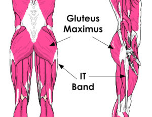
Muscle diagrams of major muscles exercised in below is an image of the outside of a normal healthy human heart diagram. Learn vocabulary, terms, and more with flashcards, games, and other study tools. These muscles include the gluteus maximus muscle (the largest muscle in the body) and the hamstrings group, which consists of the biceps femoris, semimembranosus, and semitendinosus muscles. Extension, flexion, adduction, and abduction. This is why you have to indicate which biceps you are taking about when discussing one or other of these muscles. Like the quadriceps, the hamstring muscle group also contains four separate muscles: Posterior compartment, also known as the flexor compartment; Bring one leg over the opposite thigh, and place the foot on the floor. We did not find results for: Bring museum quality art into your home or office decor with a canvas print that will never warp or sag. On the medial edge of the posterior thigh is the gracilis muscle. The groin muscles are a group of muscles situated high on the leg in the inner thigh. Posted on june 5, 2015 by admin.
This is the biggest muscle that is in the tibialis anterior. Deigram of outside leg muscles : The hamstring muscles, also known as the rear thighs, make up the backside of the upper leg anatomy. Tutorials and quizzes on muscles that act on the leg/ leg muscles (tibia & fibula), using interactive animations and labeled diagrams. Weak glutes and knee pain the biomechanics method author juli 19, 2021.
Muscles Of The Leg And Foot Classic Human Anatomy In Motion The Artist S Guide To The Dynamics Of Figure Drawing from doctorlib.info Anterior muscles of the lower leg and their functions. The hip muscles work together to carry out 4 different types of movement: This group includes the adductor magnus, adductor longus, and adductor brevis muscles, as well as the pectineus and gracilis. The gastrocnemius is the larger calf muscle, forming the bulge visible beneath the skin. It is also visible on the medial edge of the thigh from the anterior. The forearm will be parallel to the lower leg. Start studying lateral view muscles of the right leg pt 2. Your leg muscles are some of the hardest working muscles in your body.
Notice the upper leg has a biceps muscle just like the upper arm does.
Muscle anatomy gluteus 12 photos of the muscle anatomy gluteus gluteus muscle anatomy ct, gluteus muscle anatomy mri, human muscle anatomy gluteus maximus, muscle anatomy gluteus, muscle anatomy of gluteal, human muscles, gluteus muscle anatomy ct, gluteus muscle anatomy mri, human muscle anatomy gluteus maximus. Tibialis anterior, extensor digitorum longus, extensor hallicus longus, fibularis (peroneus) longus, fibu. The muscles in the hip are responsible for the movement of the hip and, by proxy, the leg. Deigram of outside leg muscles : Medial compartment, also known as adductor compartment; Deigram of outside leg muscles. However, many reflex pathways are also active in the legs and foot. This muscle is one of the ones that help. Conditions seen in this category are muscle strain, ligament and tendon strain. This is the biggest muscle that is in the tibialis anterior. Your quadricep muscles, also known as quads, consist of four muscles that compose the front of your leg; Calf muscle anatomy, calf muscle picture anatomy, lateral calf muscles, lower leg muscle anatomy, medial calf muscles, what are the two calf muscles called, what are your calf muscles called, where is your calf muscle, human muscles, calf muscle anatomy, calf muscle picture anatomy, lateral calf. Collectively referred to as the hip adductors, the groin muscles are.